Corrective Exercise Floor Glides with Leg Extension

November 14th, 2019

In this video we are bringing you a very influential exercise that does a surprisingly effective job at lower back stabilization as it challenges the mobility of the shoulders and efficiency of the the thoracic spine in extension.
As you are probably acutely aware, when working with corrective exercises and the client in pain, it is more than just the exercise, it is a thorough understanding of:

“For Whom?”

“For What?

And “When”

Insight into the biomechanics, psychological mindset of the client and social setting/implications of the movement all come into play and are critical to the client’s success.

Corrective Exercise A-P Cat and Dog Rewind

October 4th, 2019

In one of our past newsletters where we talked about myofascial mobility with rhythm, timing and amplitude, we revisited the Anterior Posterior Cat and Dog as one of the examples of applying those principles.

In this exercise, we take the A-P Cat and Dog and progress it into a surprisingly challenging core exercise. We call it the A-P Cat and Dog Rewind.

You’ll see as the clients center of mass moves forward with the change and limits of base of support, the core has to switch on in a very novel way.

The beauty of this is that the response is reflexive with no feedforward response necessary by the client or patient.

This helps us move beyond the bracing and “keep your core tight” cues that are not part of our long term goals and authentic movement.

Try it, feel it and let us know what you think.

Mobility Matters: Flexibility vs. Mobility

September 29th, 2019

via Gfycat
The terms flexibility and mobility are often used interchangeably. Although from the same “family” they are two different things. The fundamental difference is that flexibility relates to the ability of a muscle to lengthen. Mobility refers to the ability of the muscles to lengthen with control.

For example, if you can lay on your back on table and someone can push your straight leg back toward you with your knee straight so that your hip is flexed past 90 degrees, that would be your hamstring flexibility.

Then if you stood up from the table and tried to kick your leg to the same range, it is doubtful you get more than 60-80% of the range you had on the table.

That’s the difference between mobility and flexibility in its most simplified way.

Is one better than the other?

Both are important and we use both with our clientele at various times in their journey and for different objectives. But ultimately, mobility is more transferable to activities of life, work and sport. This is because mobility is a much more integrated “hardware” and “software” process. Hardware referring to the soft tissue and software referring to the nervous system.

Conversely, someone pushing on your leg (or you pulling with a strap or belt) is more of a hardware process and less of a software process then the more movement-based mobility approach.

At the end of the day, flexibility is a means to an end and not the end itself. The results of stretching can feel good and help with the compliance of our tissue.

Improvements in our range of motion (ROM) from flexibility practices can be acute (immediate but short term) and chronic (sustained improvements).

Some of the most up to date research on flexibility has shown that the most effective strategy for acute changes in ROM is self-myofascial release followed by either static or dynamic stretching. This would include foam rolling, lacrosse or tennis balls, massage sticks and other implements.

The following guide can help compare and contrast the techniques you use to feel like you have more range of motion and feel “looser”.

Self-Myofascial Release

What it is: The use of foam rollers, lacrosse or tennis balls, massage sticks and other implements in soft tissue areas of the body to produce a form of self-massage.

What it does:
Effects both the mechanical properties of the soft tissue (skin, fascia, muscles, tendons, nerves, blood vessels) and the nervous system. It reduces the resting tone of the effected muscles, leading to healthier, more compliant soft tissue.

What it doesn’t do:
Lengthen muscles or fascia

Senior woman using a foam roller for self myofascial release


Static Stretching

What it does:
Increases range of motion at a joint by lengthening the elastic component of the muscle, causing some changes in the visco-elastic properties of the muscles. Some improvements in stretching are actually attributed to the individual’s increased tolerance to the discomfort of stretching.

What it doesn’t do:
1. No clear evidence that is reduces the risk of injury prior to activity
2. May decrease force production when done prior to explosive and/or maximal effort (such as jumping, maximal lifting, etc.).

Dynamic/Active Stretching

What it does:
Increases heat and fluid exchange to the tissue. This helps with elasticity to the soft tissue. Stimulates the nervous system and thereby prepares the body for activity.

What it doesn’t do:
Provide huge gains in range of motion

Some of the most up to date research on flexibility has shown that the most effective strategy for acute changes in ROM is self-myofascial release followed by either static or dynamic stretching. The research include using foam rollers, lacrosse or tennis balls, massage sticks and other implements.

Corrective Exercise Wall Glute Bridge

August 8th, 2019

This version of the glute bridge is a great corrective exercise for teaching the body the sequencing and synchronization from the ankle to the lumbar spine that is associated with triple flexion and squatting.

It will certainly challenge dorsi flexion in a pseudo closed chain environment as the knee moves over the toes. There is feedback from the wall and load placed into the wall, but no vertical forces acting on the joint.

The body gets comfortable with relative lumbar flexion in an unload position because it has to go through that motion to get the buttocks of the floor. The beauty is both the novelty and very limited load placed on the spine. So even those with lower back pain will find this a helpful exercise as it reduces the threat.

Additionally, it helps create some separation and distraction of the tissue at the thoraco-lumbar junction. The upper part of the body is fixed on the floor as the lower part moves away, creating the distraction.

Put it to good use and tell us how you did!

How Accurate Are Your MRI Findings?

July 15th, 2019

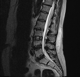
We all want the best, most accurate information we can get regarding our health. And when the doctors and therapists are not getting the results everybody wants, they will typically order imaging studies to gather more information and an “accurate” look at what is going on.

Many in the general public consider the MRI as the gold standard or best possible diagnostic available. What we must realize, however is that an MRI (magnetic resonance imaging) is an image or picture. And as such, its interpretation relies on the individual reading the image. This is generally a radiologist but may also be another medical doctor that is treating you.

The technology behind an MRI is quite amazing and is not in question. What is of concern is the accuracy of the conclusions reached and therefore the ensuing treatment plan, costs and expected outcome(s)that follow.
lumbar mri
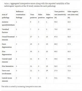
A 2017 study in The Spine Journal, wanted to look at just how accurate and consistent MRI findings would be across 10 different MRI centers. A 63-year-old women with a history of lower back pain and nerve pain down her leg received 10 MRI’s at 10 different locations over the course of 3 weeks.

What they found were very large differences in what was reported as the findings on the MRI in both false positives and false negatives (see chart from study below). Using disc herniation for example, 47.5% had a false negative or miss rate. Meaning the herniated disc was there but missed by the radiologist.

For those of us that work with a population determined to overcome their pain, the MRI is a double-edged sword. Because, as you may know from some of my past posts, the findings on an MRI do not have a one-to-one correlation with pain. There are people who have MRI’s that looked like they have been hit by a bus and have no pain. And there are people who have no clear findings on an MRI and may have significant pain. The bio-psycho-social factors that contribute to the pain experience are numerous.

What Can We Do?

Most importantly be informed and question everything. Even if you are sure the MRI findings are accurate, they don’t equate to surgery. In the absence of any medical emergency (significant muscle weakness, bowel or bladder problems), conservative treatment has shown to be as good as surgery and in several studies having even better outcomes.

At Function First, we are not doctors or physical therapists. We are exercise and movement professionals who understand the pain experience and how important it is to provide exercises that remove mechanical stress from the body and restore movement confidence through a very systematic process of program design and coaching.

When you seek help from a practitioner, you always want to be the head coach. And appreciate that passive treatments (hands on therapy, modalities) and medications may be necessary to progress you to the next step. But a comprehensive exercise program is ultimately what translates to the real-life functions you seek to participate in and enjoy.
mri variability study

Corrective Exercise-Half Frog

July 11th, 2019

This simple exercise can be quite challenging for those with a history of lower back pain, as well as the deconditioned client. The ability to disassociate the hip motion from the movement of the lower back and pelvis can be very helpful in removing stress from the lower back.

This “Level B” exercise from the PFMS is both a corrective and assessment opportunity. You will be able to see, and the client will be able to feel any control and/or range of motion differences between the two sides.

The Half Frog is also an exercise, that as a Level B allows for more intrinsic focus of attention for motor learning purposes. The client is able to concentrate on what muscles are engaging and the sensory feedback of those contractions.

Corrective Exercise-Prone Single Arm Lifts

June 14th, 2019

This corrective exercise for the shoulder and thoracic spine is significantly more challenging then it appears. A great exercise to cue the thoracic spine and scapula to better “set” their anchor points for more controlled and stable motion of the shoulder.

Three different lift angles suggested to vary the position of the gleno-humeral joint and rotation of the scapula.

How does a client achieve success with Function First?

May 20th, 2019

In this part of the interview, Anthony shares several client success stories and what the transpires during the process. Exercise is the vehicle, but there are plenty of other elements that must be in alignment as well.

Myofascial Mobility with Rhythm, Timing and Amplitude

May 10th, 2019

This video is one of the most effective underlying principles that we use to help people improve their mobility, especially when over-protective or hyper vigilant due to pain.

Using strategic movement focused on rhythm and timing with a controlled amplitude can have a profound influence on myofascial mobility.

This is not a substitute for or superior to other forms of addressing ROM and/or mobility. It is an approach to call upon, early in your intervention that may open some other opportunities for you.

This is a classic example of our “ask don’t tell” approach within the PFMS.

If nothing else, enjoy the struggle I have multiple times in this video with getting the word “expiration” to come out of my mouth 🙂

Neuroception, Relationships and Clients in Pain

May 10th, 2019

Originally written for ACE Certified by Kevin Murray

No doctor can write a prescription for creating relationships. They are hard-earned and complex undertakings, particularly with people in pain.

Part of what makes pain so distressing is its lack of predictability. Experiencing pain feeds into a negative reinforcing loop of uncertainty, up-regulating cognitive stressors such as fear, apprehension and anxiety. This often runs parallel with clients’ difficulties in regulating their emotions (Hamilton et al., 2004).

Woven into the fabric of all relationships is the principle of reciprocity. For the health and fitness professional, navigating the arena of pain and relationships requires one to become acquainted with the nervous systems role in analyzing risk and safety.

Neuroception: The Mind’s Mediator

Neuroscientist Stephen Porges coined the phrase neuroception to describe the neural mechanisms involved with subjective perception and evaluation (Van Der Kolk, 2015). Specifically, neuroception helps individuals distinguish whether a situation or individual is safe and trustworthy, or dangerous and distressing.

danger safety

To the individual experiencing pain, their unique view of the world is interpreted through a nervous system that has an altered perception or risk and safety. Every day situations can become fearful and ambiguous, often resulting in maladaptive appraisals of people who are unknown or unfamiliar.

Experiencing pain has one’s neuroceptive system on overdrive, constantly seeking out potentially threatening stimuli. This state of cognitive hypervigilance makes cultivating relationships exceptionally formidable. To combat such psychosocial stressors, successfully establishing relationships with clients in pain involves understanding the underlying mechanisms which enhance positive neuroception.

This process is governed by innate biological systems that once understood, becomes the inception of all meaningful, heartfelt and trusting relationships.

Mechanisms of the Mind

    Mirror me: Mirror you

Have you ever noticed that when someone is genuinely smiling (even if you don’t know them), you find yourself smiling back? What induces this instinctive mimicry? Why do we yawn when we see someone yawning, or wince when someone smacks their shin on a coffee table?

The neurobiological mechanisms responsible for such nonverbal imitation is regulated by highly sophisticated visuomotor neurons referred to as mirror neurons.

mirror neuron

This mirror neuron system (MNS) allows for two individuals, whether lifelong friends or two complete strangers, to simultaneously share neural activity as they attempt to decipher the meaning behind each others nonverbal gestures. The MNS is the gatekeeper of assurance and safety, escorting the manifestation of positive neuroception and is decisively involved in the emergence of all trustworthy relationships. As such, understanding the mirror neuron system’s innate bias towards familiarity and reciprocity becomes a crucial distinction with regards to clients in pain.

    Brain-to-Brain Dialog

For instance, when two people are in-sync and rapport is mutually harmonious, the MNS is fully engaged. People adopt one another’s facial expressions, hand gestures, postures. even acute motor movements without even knowing they’re doing so (Chartrand and van Baaren, 2009). This is known as automatic imitation. Interestingly, being deliberate and purposeful in the mirroring of others nonverbals (intentional imitation) can also facilitate this same mirrored neural activity between two people.

Similar neurobiological functioning ensues via verbal communication. As an illustration, when two individuals and their speech patterns converge, they adopt one another’s vocal qualities such as tone of voice, tempo of speech, even specific words and phrases. Once again, this takes place without any conscious awareness. These neural dynamics lead to mirrored neurological activity between the speaker’s brain and the listener’s brain. This is referred to as neural coupling (Stephens et al., 2010).

matching brains

In fact, have you ever experienced such high degrees of rapport where you almost knew what someone was going to say right before they said it? This is no fluke. Neural imagining via fMRI technology reveals that when two people are in-sync and engrossed socially, the delay between speech production and the listeners comprehension is so small that one can often anticipate what’s going to be said next (Hasson et al., 2011).

These anticipatory responses suggest as two individuals become acquainted with each others verbal propensities, the more attuned and mirrored their neurological activity is. Neural coupling highlights how verbal imitation can breed a sense of relatedness and commonality, ultimately nurturing the perception of safety and enhancing positive neuroception.

However, when two people are out-of-sync with their nonverbal mannerisms and verbal speech patterns, this brain-to-brain coupling vanishes (Stephans et al, 2010). When incongruencies are present, the perception of safety slowly fades and gives rise to uncertainty. If clients in pain fail to see aspects of themselves in their health and fitness professional, the more likely skepticism has the opportunity to settle in.

In-depth Analysis

The role mirroring plays in socials interactions is ubiquitous. In fact, visuomotor mimicry is so innately hard-wired that one-month-old infants display the mirroring tendencies of smiling, sticking their tongues out and opening their mouths when observing such behavior in others (Lakin et al., 2003).

As two people learn how to navigate the social complexities of interpersonal communication, what are the neurobiological intricacies involved in learning and interpreting the intended meaning of another individual’s linguistics / gestures? Let’s analyze the MNS in-action through a common example:

    Spoon Feeding and Neurobiology

As a mother brings a spoon to her infant son’s mouth for the first time, is the child aware of the next sequence required in this exchange? Does the baby open his mouth wide, accommodating for the size and shape of the spoon? Probably not.

Instead, a blank stare of bewilderment is undoubtedly written across the infant’s face. It’s not until the mother visually demonstrates the spoon-to-mouth action that the infant can comprehend what’s being asked of him.

    Sequence analysis

The infants MNS observes their parent demonstrate the action of spoon-to-mouth (intended outcome).
This creates a visuomotor representation and engages the infants own perceptual-motor circuitry.
The infant can then synthesize the visuomotor representation (action-potential) into motor execution, resulting in the reciprocation of the desired task: i.e. successfully transferring food from spoon-to-mouth for ingestion.

Here we witness the MNS and its architecture having the remarkable ability to transform passive observation, into perceptual understanding and then motor execution (Ferrari et al., 2005). Daily social exchanges such as handshakes, waving hello or goodbye, observing laughter or witnessing sadness all involve the MNS and neural coupling effects.

The mirroring of facial expressions can even result in actually adopting the emotions and moods of others (Lakin et al., 2003). This outcome is recognized as empathy, or having the capacity to understand the feelings of others and view the world through their unique perspective.

The interplay between biological and environmental factors requires more sophistication as our social surroundings increase in complexity. This makes congruent communication and mimicry as a medium for cultivating trusting relationships significant, particularly with clients in pain.

So how can you, the health and fitness professional apply these neurobiological insights with your clients in pain to enhance positive neuroception and ultimately establish relationships?

Integrating Neuroscience into Relationship Building

It’s essential to remember what distinguishes the client in pain from general clientele is their altered perception or risk and safety. Never forget, from the moment you meet your client in pain, they’re skeptically evaluating you and how you conduct yourself. As such, taking special care to remove as much uncertainty and unfamiliarity as possible becomes the primary focus. This process begins with the practice of adapting your own verbal and nonverbal mannerisms to match that of your clients.

For example, when communicating verbally, congruency is essential for positive neuroception. Suppose a client begins describing his story of musculoskeletal challenges with soft and gentle vocal qualities. He takes the time to articulate and pauses often. Attempting to mirror and reciprocate these vocal mannerisms follow the neurobiological prerequisites to manifest neural coupling

Should the client also be sitting on the edge of their seat and leaning forward, following suit and mimicking this seated posture engages the visuomotor neurons of their mirror neuron system. Intentionally integrating and reciprocating these verbal and nonverbal idiosyncrasies serves to enhance the possibility of cultivating positive neuroception.
Kevin coaching

IMAGE TAKEN FROM THE YELLOW BRICK ROAD: A 4-part framework for coaching clients in pain
The matrix of mirroring possibilities includes paying attention to your clients nonverbal features such as facial expressions, eye contact/gaze, body position and proxemics (personal space) and his or her idiosyncratic hand gestures.

Verbal and vocal aspects could encompass specific words or phrases they frequently use, paralinguistic qualities such as tone of voice, rate of speech, vocal modulation and volume, or demonstrating appropriate levels of silence should the client be reserved and introspective. Knowing which aspect(s) to mirror comes down to actively listening and observing the uniqueness of each clients’ communication tendencies.

As clients in pain begin experiencing coherence and familiarity in your communication conduct, their skepticism is superseded with impressions of trust and certainty. Their perception of safety and assurance increases as positive neuroception begins planting its roots.

And while the genesis of cultivating relationships varies from one individual to the next, attempting to enter each client’s world and speak their language helps to nurture the inception of meaningful, heartfelt and trusting relationships with your clients in pain.